Instant pathology: Transforming cancer diagnostics
Flash Pathology empowers researchers and clinicians with advanced imaging solutions. Our groundbreaking label-free technology enables real-time visualization of unprocessed tissue, offering precision and speed like never before. Revolutionize biopsy analysis and surgical margin assessment with clarity you can trust.
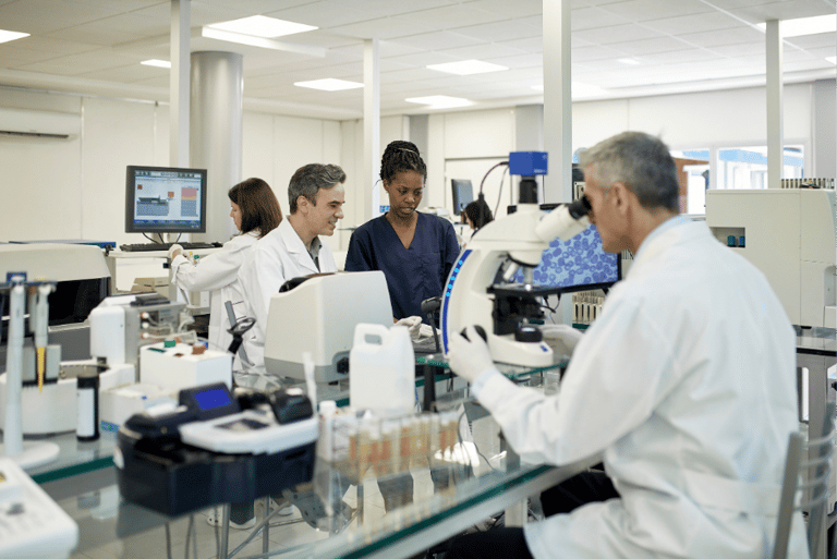
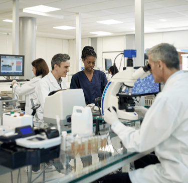
Fast, comprehensive tissue analysis
Today's challenges in pathology
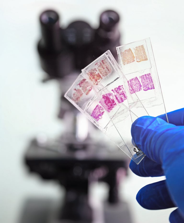
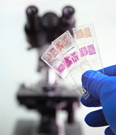
Advancements in traditional H&E staining have improved processing times, but the overall turnaround for results often still takes several days. This delay can be further exacerbated when new biopsy samples are required due to inadequate representation of the initial specimen.
Delays in diagnostics and treatment
The inability to perform intraoperative biopsy analysis leads to a high rate of re-interventions, both for additional tissue sampling and follow-up surgeries. This not only increases patient risk but also imposes a significant cost burden on hospitals.
High costs and increased risk of re-interventions
Traditional histology methods, such as H&E staining of paraffin-fixed or frozen tissue sections, render samples less suitable for subsequent molecular or genetic analysis. This limitation prevents pathologists from accessing critical information that could guide more precise diagnoses and optimize patient treatment plans.
Increased need for high-precision analysis
The future of pathology, today
Traditional H&E slide preparation involves 14 meticulous steps, from tissue excision to fixation and staining. While highly efficient, bypassing this process could significantly reduce costs for healthcare systems and shorten the time to diagnosis for patients.
Flash Pathology’s innovative technology eliminates the need for fixation, slicing, or staining. Our system allows tissue to be analyzed in its fresh, unprocessed state—saving valuable time and resources while accelerating diagnostic workflows.
Streamlined tissue analysis
Get it right the first time
Even with advancements in imaging and robotic assistance, biopsy samples often fail to capture the required cells for histopathological analysis. This leads to incomplete or failed lab results, repeat biopsies, and delayed diagnoses.
Flash Pathology provides reliable, real-time imaging that helps ensure samples are representative the first time, reducing the need for repeat procedures and expediting patient care.
Real-time insights
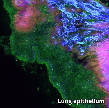
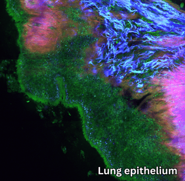
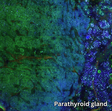
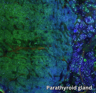
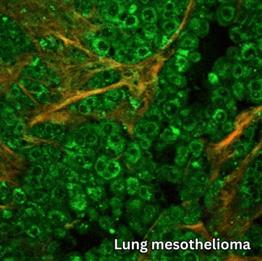
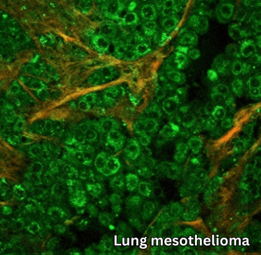
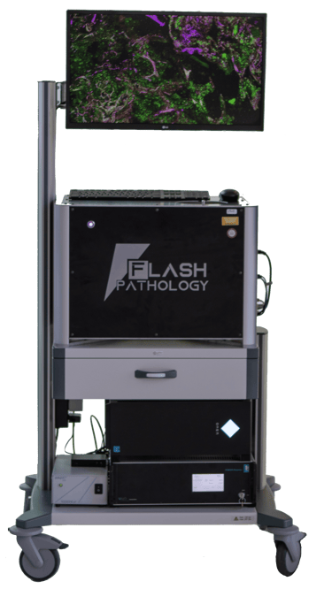
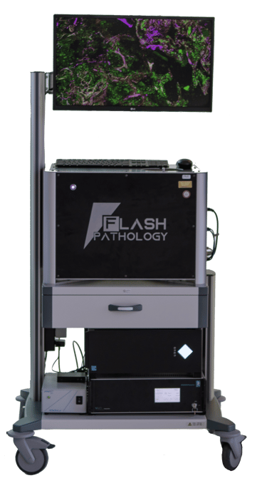
Using ultrashort infrared laser pulses, Flash Pathology’s technology generates up to four distinct signals, each revealing unique tissue characteristics. The result is high-resolution microscopy images packed with rich diagnostic information.
These images are immediately interpretable, offering diagnostic clarity even before applying computational analysis for deeper insights. Additionally, the technique provides optical sectioning of the tissue in thin slices, which can be combined to create detailed 3D visualizations of the sample.
Flash Pathology B.V.
Paasheuvelweg 3
1105 BE Amsterdam
The Netherlands
Contact
Menu
Technology
At Flash Pathology, we are committed to empowering you with advanced tools to visualize and understand the intricate details of tissue samples.
Our products are designed and developed under ISO 13485 certified processes