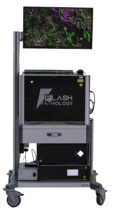
Flash Scanner FS4-R
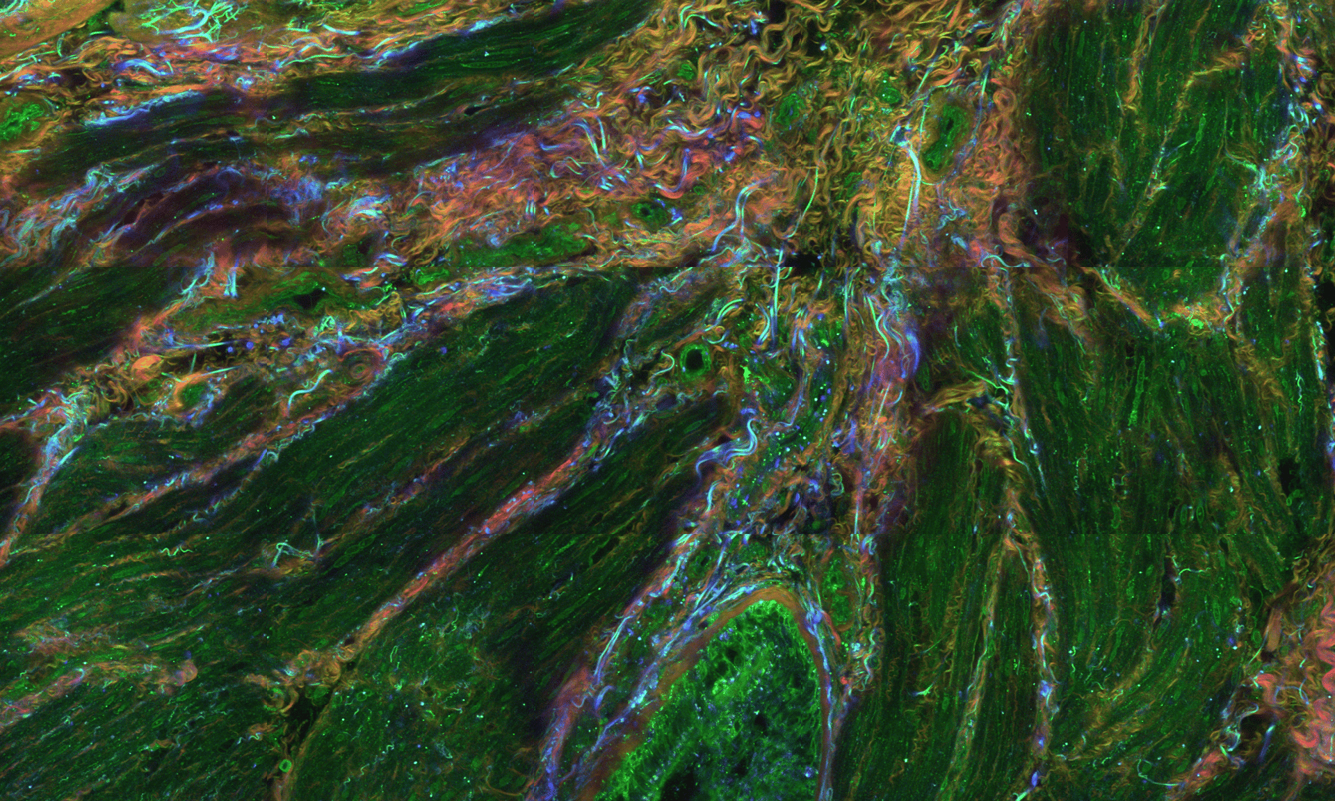
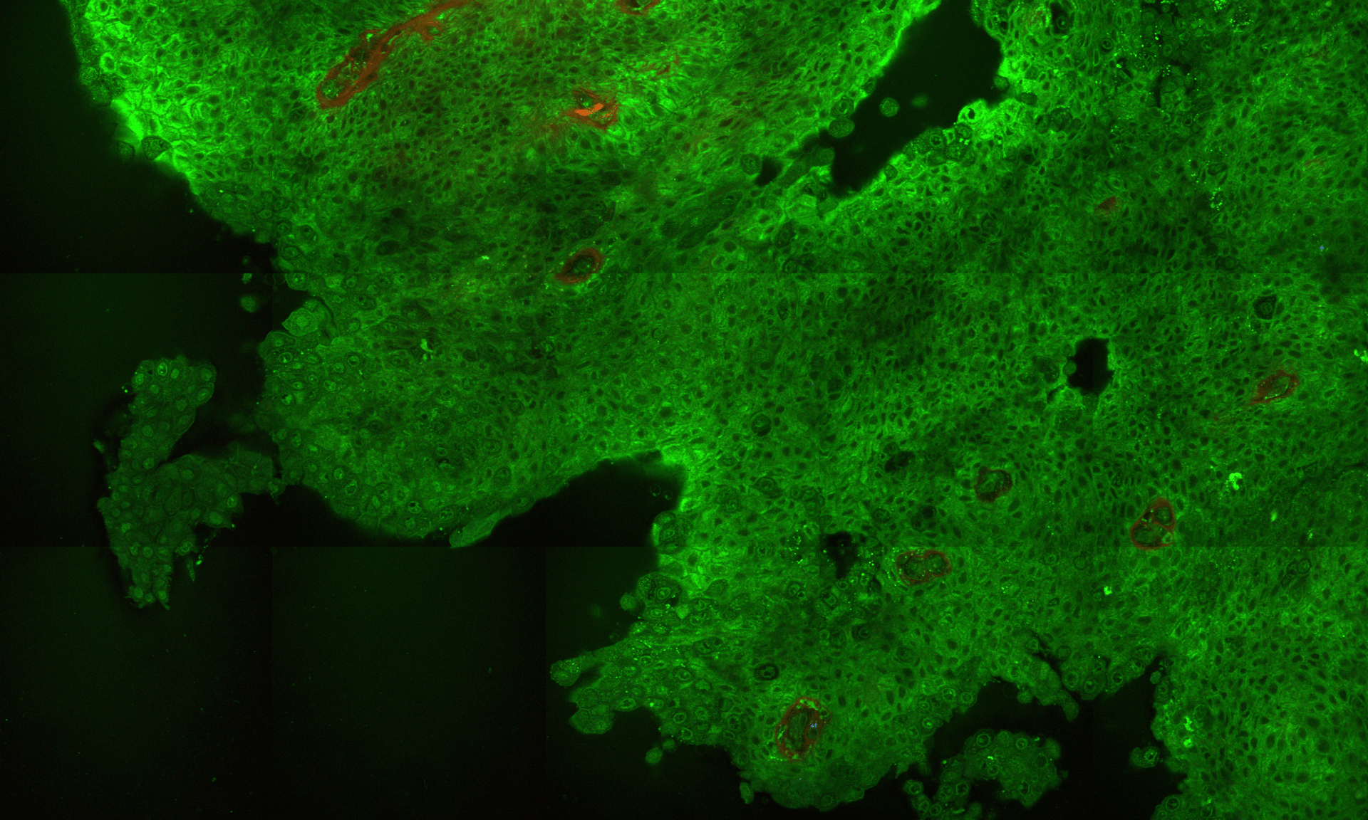
The Flash Scanner FS4-R is a state-of-the-art microscope that harnesses higher harmonic generation (HHG) to deliver rapid, label-free imaging of excised tissue, revealing intricate details at the cellular and subcellular levels. Whether examining the spatial architecture of cells in the tumor microenvironment, studying the effects of drugs on cellular behavior, or gaining insights into tissue development without invasive labeling, the Flash Scanner FS4-R reveals cellular and histological hallmarks with unmatched speed and precision, marking a significant advancement in tissue analysis.

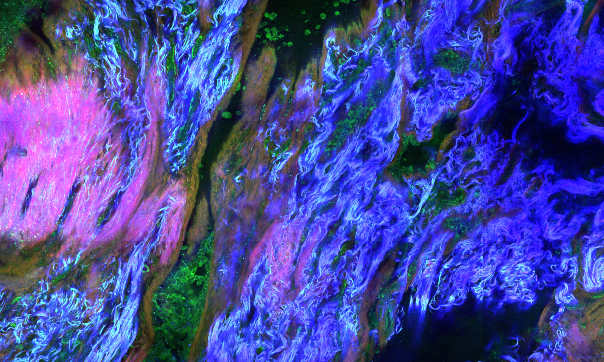
>> Subcellular resolution
Reveal key cellular and histological hallmarks using four distinct imaging modalities.
>> Label-free imaging
Visualize tissues without the use of labels or dyes, preserving samples for further analysis.
>> 3D tissue visualization
Generate 3D images using automatic optical sectioning - no sample slicing required.
>> Quick turnaround
Minimize specimen preparation time and evaluate your samples in just minutes.
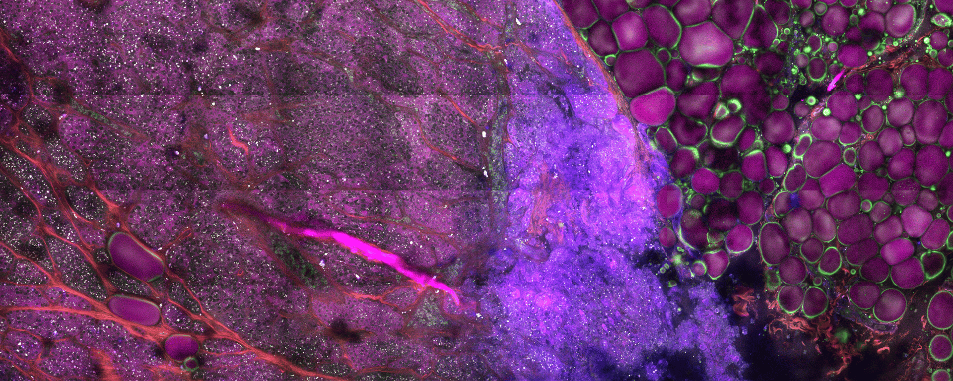
>> Publication-ready images
Capture vibrant, high-quality images that effectively convey the story behind your data.
Frequently asked questions
How does the Flash Scanner FS4-R work?
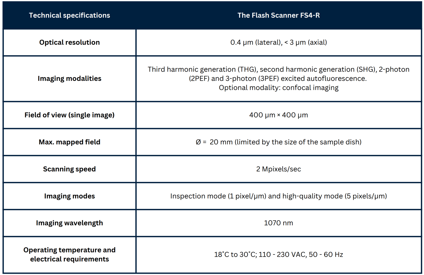
The Flash Scanner FS4-R is an imaging platform designed for rapid, label-free histological assessment of unprocessed tissue samples. The functionality of the Flash Scanner is based on the detection of nonlinear multiphoton signals, including higher harmonic generation (HHG) signals, such as second and third harmonic generation (SHG and THG), as well as two-photon and three-photon excited autofluorescence (2PEF and 3PEF). These imaging modalities provide complementary information on both tissue architecture and cell morphology, allowing to successfully discriminate between different types of tissues.
What tissue structures can be visualized with the Flash Scanner?
The Flash Scanner FS4-R offers four unique imaging modalities – second harmonic generation (SHG), third harmonic generation (THG), two-photon excited autofluorescence (2PEF), and three-photon excited autofluorescence (3PEF):
SHG: Captures non-centrosymmetric molecules and structures, like collagen fibers, microtubules, and myofilaments.
THG: Visualizes cellular structures with distinct refractive indices, such as cell membranes, lipid droplets, and myelin sheaths.
2PEF and 3PEF: Detect the presence of intracellular molecules, such as flavins, NAD(P)H, retinol, and tryptophan, as well as extracellular structures, including elastin and collagen fibers.
Can the Flash Scanner FS4-R image tissue samples without any preparation or processing?
Yes, the Flash Scanner FS4-R can image fresh tissue samples without the need for staining, labeling, or other forms of tissue processing. Higher harmonic generation directly utilizes the optical properties of the tissue, allowing for high-resolution imaging while preserving the sample’s natural structure and composition.
Can the tissue samples imaged with the Flash Scanner FS4-R be used for further analysis?
Yes, the tissue samples imaged with the Flash Scanner FS4-R can be used for further analyses, such as histology, molecular studies, or other imaging techniques. The Flash Scanner utilizes non-invasive, label-free imaging modalities, such as higher harmonic generation and multiphoton excited autofluorescence, therefore the samples remain intact and undamaged.
Flash Pathology B.V.
Paasheuvelweg 3
1105 BE Amsterdam
The Netherlands
Contact
Menu
Technology


At Flash Pathology, we are committed to empowering you with advanced tools to visualize and understand the intricate details of tissue samples.


Our products are designed and developed under ISO 13485 certified processes
